Lumpy skin disease: Differential diagnosis
Learn more about diagnosing LSD 29 June 2023
 2 minute read
 By:
By: Part of Series:
Editors note: The following content is an excerpt from Lumpy Skin Disease: a field manual for veterinarians which is designed to enhance awareness of lumpy skin disease and to provide guidance on early detection and diagnosis for private and official veterinary professionals (in the field and in slaughterhouses), veterinary paraprofessionals and laboratory diagnosticians.
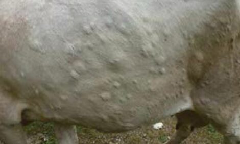
Severe cases of LSD are highly characteristic and easy to recognize. But early stages of infection and mild cases may be difficult to distinguish even for the most experienced veterinarians, requiring a laboratory confirmation. Samples should be collected from all suspected animals and tested using fast and highly sensitive PCR methods to differentiate true cases. The following diseases may be considered as a differential diagnosis for LSD:
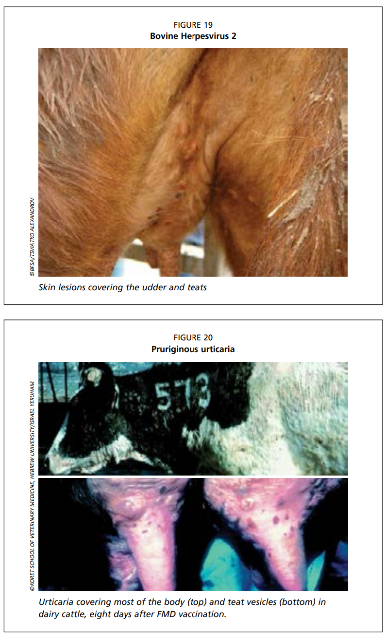
- Pseudo lumpy skin disease/Bovine herpes mammillitis (bovine herpes virus 2) (Fig. 19): dermal lesions may look like those caused by LSDV, but are more superficial and the course of the disease is shorter and less severe. The disease can be ruled out by detecting LSDV by PCR.
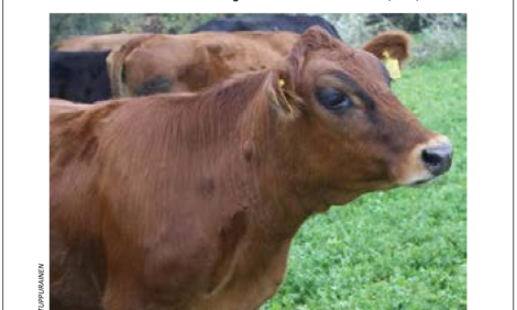
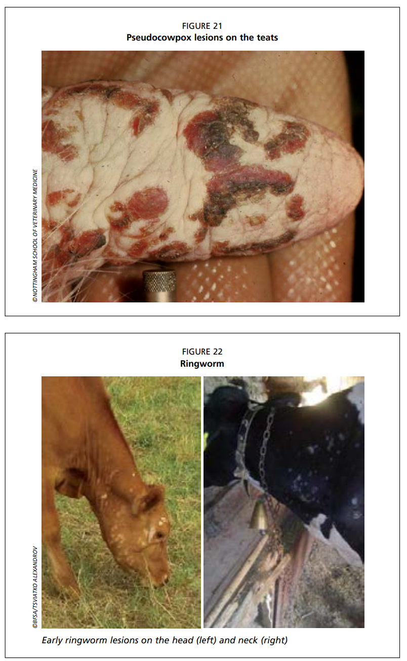
- Insect bites, urticaria, and photosensitisation: dermal lesions may look like those caused by LSDV, but are more superficial and the course of the disease is shorter and less severe (Fig. 20). The disease can be ruled out by detecting LSDV by PCR.
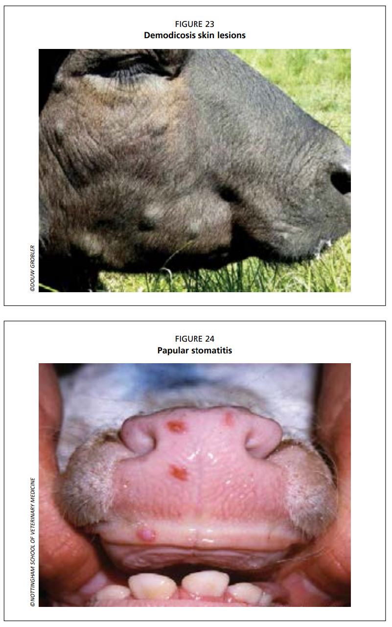
- Pseudocowpox (Parapoxvirus) (Fig. 21): lesions occur only on the teats and udder. The disease can be ruled out by detecting LSDV by PCR.
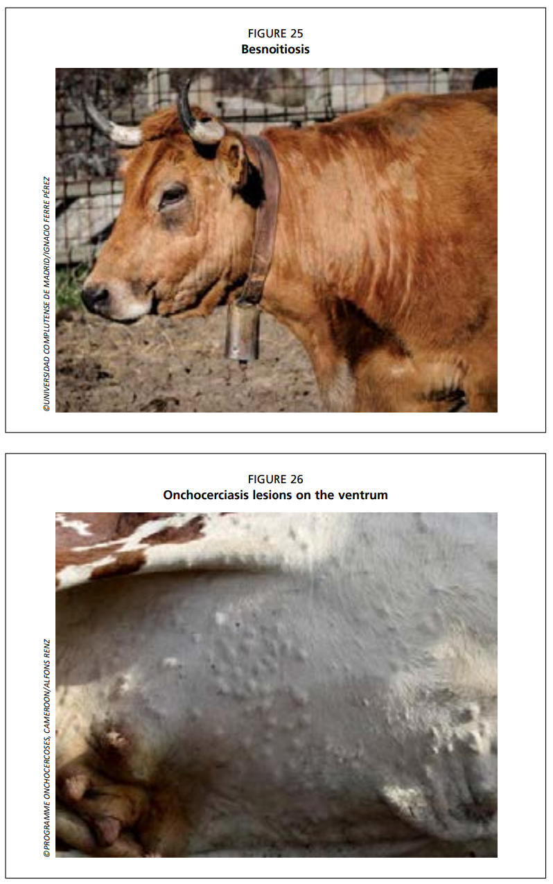
- Dermatophytosis (Fig. 22): early ringworm lesions, more superficial, clearly different, non-ulcerative surface structure of the ringworm lesion.
- Demodicosis (Fig. 23): dermal lesions predominantly over withers, neck, back, and flanks, often with alopecia present. The disease can be ruled out by detection of mites using skin scrapings.
- Bovine papular stomatitis (Parapoxvirus) (Fig. 24): lesions occur only in the mucous membranes of the mouth. The disease can be ruled out by PCR testing.
- Besnoitiosis (Fig. 25): lesions often occur in scleral conjunctiva, and dermal lesions may exhibit alopecia with thick and wrinkled skin. The disease can be ruled out by detecting LSDV by PCR.
- Onchocerciasis (Fig. 26): dermal lesions most likely at ventral midline. The disease can be ruled out by PCR. In addition, live, attenuated LSDV vaccines may cause mild adverse reactions in cattle which resemble clinical LSD (see pp. 37-40 for currently available vaccines).
Reference:
Tuppurainen, E., Alexandrov, T. & Beltrán-Alcrudo, D. 2017. Lumpy skin disease field manual – A manual for veterinarians. FAO Animal Production and Health Manual No. 20. Rome. Food and Agriculture Organization of the United Nations (FAO). 60 pages.
Headline image courtesy of ©BFSA/Tsviatko Alexandrov