


Udder Examination
Dairy farmers are aware of the changes seen in clinical mastitis. However sub-clinical mastitis is by its very nature more difficult to detect.Most UK large animal vets and farmers do not routinely palpate udders. Many European vets and farmers find udder palpation a valuable tool.
A proportion of chronic high cell count cows will have discernable abnormalities such as the formation of abscesses, scar tissue and irregular nodules in the udder tissue. These animals are unlikely to respond to treatment and constitute a risk to other cows in the herd by acting as a reservoir of infection. The best option is to milk these cows last or preferably remove them from the herd.
Palpation technique
Palpate the udder immediately after milking when it is flaccid.Skin
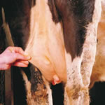 Grasp the udder skin between the forefinger and thumb; it should be pliable and easily separated from the underying tissue. If not it is likely that there is
oedema (swelling) of the udder associated with infection or recent calving.
Grasp the udder skin between the forefinger and thumb; it should be pliable and easily separated from the underying tissue. If not it is likely that there is
oedema (swelling) of the udder associated with infection or recent calving.
Udder tissue
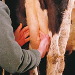 Palpate each quarter with
both hands by placing
one hand on the inner
side and the other on the
outer side. Deep palpate
by pressing the finger tips
towards each other and
gradually work the hands
towards the bottom of each
quarter. The udder tissue should
have a fine grain. A coarse grain that still feels soft
should respond to treatment. Cows with a coarse
grained, harder udder or those with any lumps or
nodules are less likely to respond well.
Palpate each quarter with
both hands by placing
one hand on the inner
side and the other on the
outer side. Deep palpate
by pressing the finger tips
towards each other and
gradually work the hands
towards the bottom of each
quarter. The udder tissue should
have a fine grain. A coarse grain that still feels soft
should respond to treatment. Cows with a coarse
grained, harder udder or those with any lumps or
nodules are less likely to respond well.
Lymph nodes
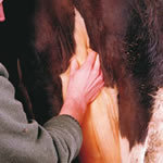 The most obvious
mammary lymph nodes
lie above each hind
quarter at the base of the
udder between the hind
limbs in what is known as
the perineal region. These
lymph nodes are normally
inapparent but will enlarge and
feel nodular with infection.
The most obvious
mammary lymph nodes
lie above each hind
quarter at the base of the
udder between the hind
limbs in what is known as
the perineal region. These
lymph nodes are normally
inapparent but will enlarge and
feel nodular with infection.
If this technique is done regularly on high cell count cows the vet and farmer can select the animals most likely to respond to therapy. Cows with a poor chance of recovery can be excluded from treatment programmes. This will result in a cost effective approach to curing high cell count cows.
Farmers Guides
- Optimising the milking routine
- Basic mastitis types and control
- Probability of cure following intramammary antibiotic treatment
- Improving udder health around
drying off - Udder Examination
- California Mastitis Test
- Technique for infusion of a combination of dry cow antibiotic and OrbeSeal®
- Sterile milk sampling for bacteriology
- Collecting and transporting samples
- Risks of feeding dump milk to calves