Anatomy of the Cow’s Reproductive Tract
By Dr. R.W. Prange and Dr. R.T. Duby University of Massachusetts and published by West Virginia University Extension Service. Successful reproduction on modern dairy farms requires an understanding of reproductive processes of the dairy cow and a working knowledge of the anatomy or parts of a cow’s reproductive tract. 11 June 2007
 12 minute read
This knowledge can be useful in identifying and correcting many situations leading to poor reproductive efficiency.
Except for the vulva, all parts of the reproductive tract are located within the body of the cow. Parts (Fig. 1) encountered as one proceeds into the reproductive tract include the vestibule, vagina, cervix, uterus, oviducts and ovaries. The internal parts are located beneath the rectum, which allows rectal palpations of the tract to be done easily. The uterus, oviducts, and ovaries are attached to a ligament and suspended in the cow’s pelvic area. This suspension allows these organs to move freely in the pelvic canal and into the body cavity, providing space to accommodate a growing fetal calf.
The Reproductive Tract
Vulva
The vulva (Fig. 1) is the external part of the reproductive tract. The thickened folds of skin of the structure are sensitive to changes in estrogen, the hormone (Fact Sheet IRM-2) responsible for estrus (heat). Swelling and redness of the vulva, due to increased blood flow, can be useful in estrous detection when coupled with other signs.
Vestibule
The vestibule (Fig. 1) is a part of the reproductive tract shared with the urinary system. It is approximately 4 inches long. Openings from the urinary bladder and a blind sac located below the opening of the urethra called the suburethral diverticulum are located on its floor. Dairy producers and Al technicians can prevent insertion of an inseminating rod into these openings, which could result in injury or insemination failure, by knowing their location.
Vagina
The vagina is located between the opening to the bladder and the cervix. Approximately 8 inches long (Fig. 1), it is the site of semen deposition during natural service. The vagina also serves as an unrestrictive passageway for the calf at time of birth. One important function of the vagina is as a line of defense against invasion by bacteria. The epithelium of the vagina secretes fluids which combine with cervical fluids to inhibit growth of undesirable bacteria.
Protection from infections may not be sufficient when unsanitary housing conditions are prevalent, or dirty inseminating equipment is used. As a result, vaginal infections can be a problem. In addition, pooling of urine in the vagina adjacent to the cervix can cause infertility in some older cows.
Cervix
The cervix (Fig. 2) is a unique structure within the reproductive tract. It is 4 to 5 inches long and 1 to 2 inches in diameter and lies between the vagina and uterus. This structure is designed to restrict access to the uterus. The area around the opening of the cervix actually protrudes back into the vagina. This protrusion deflects such things as inseminating rods away from the cervical opening if care is not taken during insemination.
Also, the walls of the cervix are thick and dense in comparison to the walls of the vagina. Three or four ridges or rings within the body or the cervix, called annular folds, can be distinguished by rectal palpation (Fig. 2). The folds must be manipulated rectally while an inseminating rod is passed through to the uterus.
The cervix has important functions. The anterior cervix may serve as a site for semen deposition during artificial insemination (Al). This occurs on services where the cycle length is not 21 days and pregnancy from a previous service is possible. Whether by deposition following Al or by migration from the vagina after natural service, the cervix acts as a reservoir for semen. The cervix provides a favorable environment for sperm survival.
Secretions of the cervix are usually thick, but these fluids thin at the time of estrus to facilitate transfer of sperm to the uterus. Some of the mucus may be seen as discharge from the vulva around the time of estrus. The cervix, or fluids of the cervix, act as a physical barrier and protect the uterus from any foreign material or bacteria during pregnancy. A thick plug forms in the canal of the cervix and blocks access to the pregnant uterus. Accidental rupture of this plug by insertion of an inseminating rod can result in abortion.
 |
 |
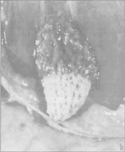
| Fig. 1. Parts of a cow’s reproductive tract. | Fig. 2. Cervix, body of uterus and beginning of uterine horns of the reproductive tract of the dairy cow. The cervix and a small portion of the uterus has been cut open. Note the thick folds of the cervix. The body of the uterus is less than 2 inches long before it divides (at arrow) into the uterine horns. |
Uterus
The uterus (Fig. 1) consists of a “body” and two “horns”. It is attached to the broad ligament and suspended within the pelvic cavity and posterior portion of the body cavity. The body of the uterus is adjacent to the cervix. In a non-pregnant state it extends less than 2 inches before it divides into two separate horns (Fig. 2).
The uterine body is the major site of semen deposition during Al. If the tip of the inseminating rod is inserted too far into the uterus, semen is deposited in only one of the uterine horns (Fact Sheet IRM-12). If the egg was released from the ovary on the other side, there is little chance that sperm and egg would unite. Remember, the body of the uterus is less than 2 inches long and caution must be used to correctly deposit semen into this region.
The uterus has many functions. Its walls are composed of several layers of muscle which aid in transport of sperm to the oviduct following insemination and in expulsion of the calf at birth. Certain glands within the walls of the uterus secrete a fluid, uterine milk, which provides nutrients to the developing embryo before and after its attachment to the uterine wall.
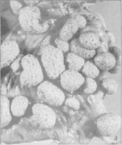
The uterus also develops the maternal side of the placenta to nourish and protect the developing fetus. Its surface contains many specialized areas called caruncles (Fig. 3). Cotyledons of the fetal placenta interlock (Fig. 3) with caruncles on the uterus to provide a passageway for the exchange of nutrients and waste between fetus and cow. After calving, if the caruncles and cotyledons fail to unlock, the placenta cannot be expelled and a retained placenta (Fact Sheet IRM-21) results.
 |
 |
| Fig. 3. Lining of uterus (a) showing caruncles on its surface. The caruncles interlock with the cotyledons (b) on the fetal placenta to provide a passageway between the cow and fetus for transfer of nutrients and waste products. Retained placentas result when the cotyledon and caruncle fail to separate after calving. | |
Oviduct
The oviducts (Fig. 1) are approximately 10 inches long, 1/4 inch in diameter and lie between each ovary and tip of the adjacent uterine horn. The ovarian end of the oviduct is funnel shaped and called the infundibulum. The infundibulum catches the egg as it is released from the ovary at ovulation and moves it to the enlarged upper end of the oviduct called the ampulla. Fertilization occurs here within 12 hours of ovulation. After fertilization, the fertilized ovum is transported to the uterus in a process requiring 3 to 4 days.
Ovaries
The ovaries are the primary reproductive organ of the female. In a dairy cow, each ovary is approximately 1.5 inches long and 3/4 inch in diameter (Fig. 4). The ovaries are suspended from the broad ligament near the end of the oviduct and lie near the tips of the curved uterine horns. Their function is to produce the egg or ovum and hormones involved in regulating the estrous cycle and pregnancy.
The ovaries contain thousands of ova. These are produced by the embryo prior to birth. While the potential to collect hundreds of ova from a cow exists, only one ovum is usually released during each estrous cycle. When more than one ovum is naturally released it can lead to multiple births—an undesirable event because of freemartinism. However, superovulation, or the production of several ova following injection of hormones such as pregnant mare’s serum gonadotropin (PMSG) or follicle stimulating hormone (FSH), is an essential element in embryo transfer.All ova are surrounded by a special layer of cells in the ovary. The growth of these cells produces blister-like structures, called follicles, that are visible on the surface of the ovary (Fig. 4).
These develop continuously throughout the life of the cow and the vast majority regress without releasing the ova. Development of ovulatory follicles begins at puberty. As the follicle enlarges, it appears as a large blister on the surface of. the ovary and can be easily detected by rectal palpation. This phase of activity is culminated by the release of ova from the follicle along with the follicular fluids.
Following ovulation, the walls of the follicle collapse and develop into the corpus Iuteum (CL) or yellow body (Figs. 4,5). The CL reaches its maximum size 10-12 days after ovulation and is the dominant structure on the ovary. If a pregnancy does not result, the CL regresses 3 to 4 days prior to the next ovulation. However, the presence of an embryo in the uterus prevents this from happening.
Development of the follicle and subsequent formation of the CL are associated with the production of estrogen and progesterone, respectively. Estrogens are produced by the cells lining the wall of the follicle and are responsible for changes in behavior as well as altering the production of fluids by the vagina, uterus and cervix. In addition, estrogens also trigger the release, from the pituitary gland, of the hormone responsible for ovulation, Iuteinizing hormone (LH).
As a result of these synchronized events, the cow comes into estrus, can be mated, the fluids of the tract provide a favorable environment for survival of the sperm and ova, and ovulation occurs at the time when sperm will be available to cause fertilization.
 |
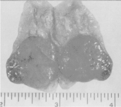 |
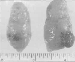
| Fig. 4. Ovaries of the dairy cow. Note the blisterlike follicle on the left ovary. The large structure on the right ovary is a corpus Iuteum. It is yellowishorange. A fully developed corpus Iuteum will occupy a major portion of the surface and body of the ovary. | Fig. 5. Ovary cut to show relative size of mature CL. Most of the CL is located within the body of the ovary. This structure produces and secretes progesterone, the hormone responsible for maintenance of pregnancy. |
Associated with ovulation is the transformation of the follicle wall into the CL under the influence of LH. The CL begins to produce progesterone which is required for maintenance of pregnancy. Progesterone acts upon the lining of the uterine wall to prepare it for subsequent attachment of the embryo. In addition, progesterone and low levels of estrogen prevent resumption of normal cyclic activity and allow for maintenance of pregnancy (Fact Sheet IRM-2).
Another important function of the CL is the production of a hormone called relaxin. Relaxin relaxes the cervix and suspensory ligaments in the pelvic region prior to calving, producing the “springer” look. This relaxation of the cervix is essential for the successful delivery of a new calf. Induction of parturition with oxytocin prior to relaxation of the cervix may result in damage to the uterus because the cervix may not relax sufficiently to allow for passage of the calf.
Pregnancy and the Reproductive Tract
Major changes in the ovaries, uterus and cervix occur during pregnancy. The presence of the CL on the ovary during pregnancy prohibits development of mature follicles. The uterus enlarges as do the sites of embryo attachment, the caruncles. In the non-pregnant cow these structures are approximately 1/2 inch in diameter, but are 2-3 inches in diameter at calving. A thick mucus plug forms in the cervical canal and is thought to protect the uterus from infections. The walls of the vagina and vulva are dry and white because of the lack of estrogens.
The relative position of the reproductive tract within the pelvic arch also changes. As the embryo increases in size, the pregnant horn begins to drop over the rim of the pelvis into the body cavity and displaces the intestines. As it does so it stretches the ligaments and pulls the ovaries along with it. As a result, palpation of the ovaries during pregnancy may be difficult. The uterine horn containing the embryo enlarges at a more rapid rate than the “non-pregnant” horn which accommodates a portion of the placenta.
The birth process may be divided into two parts: the delivery of the calf and the subsequent delivery of the placenta or after birth in a process referred to as “cleaning.” It must be remembered that the attachment sites of the placenta in the uterus consist of the maternal contribution, the caruncle, and a fetal side called the cotyledon. During pregnancy these tissues interlock with each other forming tight attachments (Fig. 3).
When the calf is born, the fetal placenta loses its source of nourishment, blood pumped by the calf heart. The loss of nutrients coupled with changes in the caruncle brought about by decreases in progesterone lead to a loosening of the attachment sites. The placenta is then passed by strong uterine contractions illicited by prostaglandins from the uterus and oxytocin released by nursing or milking.
Failure of the release mechanisms, due to hormonal imbalance, nutritional imbalances (Vitamin E & Selenium) or infections that cause swelling of the tissues, results in a retained placenta (Fact Sheet IRM-21). After “cleaning” occurs it takes the uterus 30-40 days to return to its non-pregnant size and condition. Discharges during this period are due to the repair of the uterine tissues and should be of little concern unless they contain pus. This would indicate the presence of a local infection that would require treatment (Fact Sheet IRM-22).
Abnormalities of the Reproductive Tract
Abnormalities may be classified as structural or functional and are estimated to account for 10-20% of infertility in dairy cattle. A structural abnormality could be the result of abnormal embryonic development while functional abnormalities could be due to hormonal imbalances.
The most familiar structural abnormalities are seen in the freemartin. The birth of a heifer co-twin to a bull results in abnormal development of the heifer’s reproductive tract. This condition results when the placentas of the embryos unite during pregnancy, allowing the two embryonic circulations to combine. As a result, a substance responsible for organizing the male reproductive system crosses into the female and inhibits the development of the ovaries.
In addition, the development of the oviducts, uterus, cervix and part of the vagina is blocked by a substance produced by the developing testis of the male. The degree of inhibition is related to the stage at which the placentas joined. The earlier the fusion, the more complete the inhibition. Other abnormalities such as two cervices, absence of one uterine horn, or blockage of the oviducts also occur.
Functional abnormalities such as cystic ovaries (Fact Sheet IRM-25) or infections in the oviducts or uterus (Fact Sheet IRM-22) leading to the distention with pus are also common. While theories exist to explain the causes of these conditions, treatment consists of hormone or antibiotic therapy.
Summary
The reproductive tract of a cow is composed of the vulva, vestibule, vagina, cervix, uterus and ovaries. The ovaries, under control of the hormones FSH and LH from the pituitary, mediate events of the reproductive cycle and reproductive tract through secretion of ovarian hormones, estrogens, progesterone and relaxin. The ovaries also release ova which carry the maternal genes. Structural abnormalities such as freemartinism, double cervix, etc. can impede reproduction. A lack of understanding of the anatomy of the reproductive tract can lead to poorer conception rates and lower reproductive efficiency.
Trade or brand names are mentioned only for information. The Cooperative Extension Service intends no endorsement nor implies discrimination to the exclusion of other products which also may be suitable.January 2007